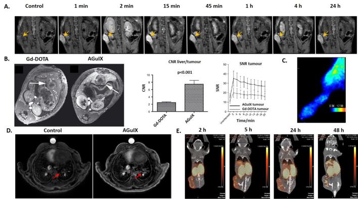
Figure 4.
(A) T 1 weighted MRI of a pancreatic tumour bearing mouse after i.v. administration of AGuIX NPs. Yellow arrows indicate tumour localization. (B) T 1 weighted image of hepatic colorectal cancer metastasis after i.v. administration of molecular agent (Gd-DOTA) and AGuIX NPs. Comparison of contrast-to-noise ratio and signal-to-noise ratio for Gd-DOTA and AGuIX NPs in tumour. (C) In vivo SPECT imaging in the paw of a swarm rat chondrosarcoma orthotopic model after i.v. administration of radiolabelled 111In AGuIX NPs. (D) Ultrashort echo-time MRI axial slices of a lung tumour bearing mouse before and after i.v. administration of AGuIX NPs. (E) PET/CT imaging of a 4T1 tumour bearing mice after i.v. administration of radiolabelled 64Cu AGuIX NPs. Adapted from,35,46–48. i.v., intravenous; NPs, nanoparticles; SPECT, single photon emission computed tomography.

