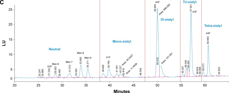
Figure 3.
Chromatograms of the N-linked glycan charge separations for (A) rAHF-PFM; (B) rFVIII-FS; and (C) BAY 81–8973.
Notes: Separation of the neutral, mono-, di-, tri-, and tetra-sialylated N glycans are shown in their respective retention time windows. Major glycan structures are annotated.
Abbreviations: LU, luminescence units; rAHF-PFM, antihemophilic factor (recombinant) plasma/albumin-free method; rFVIII-FS, sucrose-formulated recombinant factor VIII.


