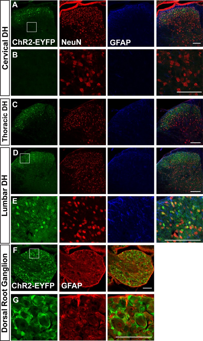
Fig 5.
A. Images of cervical dorsal horn (DH) (A&B), thoracic DH (C), lumbar DH (D&E) and dorsal root ganglion (F&G) of adult Ai32/Ai32 mice stained with anti-GFP, anti-NeuN, and anti-GFAP antibodies. B shows a higher magnification view of ChR2 non-expressing region boxed in A. E and G show higher magnification views of regions boxed in D, F respectively. n = 3 mice, scale bars = 100 μm.

