. 2019 Mar 13;8:e45100. doi: 10.7554/eLife.45100

Figure 1. Generation of RPEΔAbca1;Abcg1 mice.

(A) IF staining for ABCA1 (yellow), ABCG1 (violet) and the RPE apical marker EZR (white) in retinas of 2-months-old wt mice. Lower panels show magnification of the RPE layer. Nuclei were counterstained with DAPI (blue). Ch: choroid; RPE: retinal pigment epithelium; ONL: outer nuclear layer; INL: inner nuclear layer; GCL: ganglion cell layer. (B) Cre mRNA levels were measured by semi-quantitative real-time PCR in neural retinas and eyecups (RPE/choroid) from 2-months-old RPEΔAbca1;Abcg1 mice. Shown are data from individual samples and means ± standard deviations (SD, N = 4). Statistics: Student’s t-test; ***: p<0.001. (C) IF staining for CRE (red) in retinal sections from 2-months-old Ctr and RPEΔAbca1;Abcg1 mice: white arrowheads indicate CRE-positive nuclei in the RPE of mutant mice. Nuclei were counterstained with DAPI (blue). Note the non-specific signal in the inner retina. Representative pictures of N = 6 mice. (D) Detection of CRE-mediated excision fragments in Abca1 and Abcg1 (Abca1/Abcg1 exc) by conventional PCR on genomic DNA from eyecups and neural retinas of Ctr and RPEΔAbca1;Abcg1 mice (N = 3). For this picture, animals showing heterozygous deletion of Abca1/Abcg1 in ear biopsies (see ‘Materials and methods’) were excluded in order to detect excision truly due to CRE expression in the eye. PCR for the floxed sequences (Abca1/Abcg1 flox) was performed as positive control. Shown are PCR products run on a 2% agarose gel and visualized with ethidium bromide. Note the lack of the excised fragment in the neural retina. M: DNA size marker, indicated fragment sizes are shown in base pairs (bp).

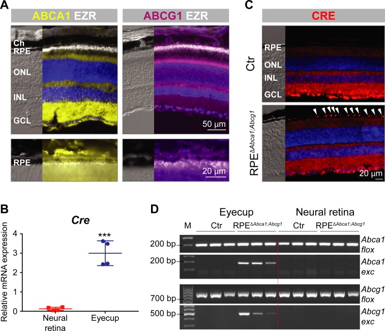
Figure 1.

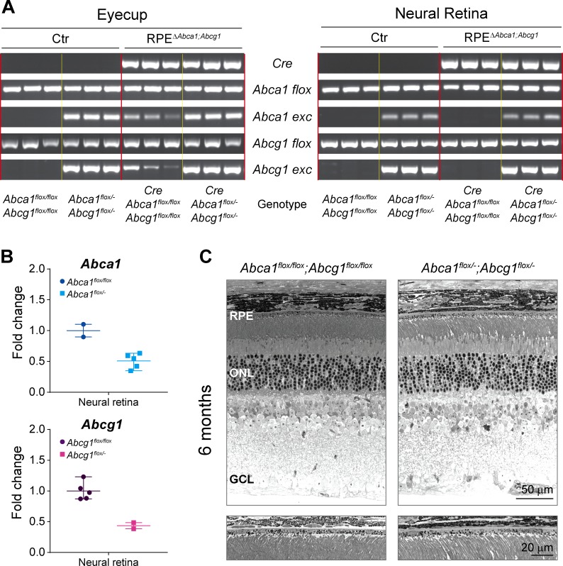
Figure 1—figure supplement 1. Genotyping and definition of the mouse models.

Figure 1—figure supplement 1.

(A) PCRs for Cre transgene, Abca1 floxed (flox) and excised (exc) alleles, and Abcg1 floxed and excised alleles were performed on genomic DNA extracted from eyecups and neural retinas of 2-months-old Ctr and RPEΔAbca1;Abcg1 mice. Shown are PCR products run on a 2% agarose gel and visualized with ethidium bromide. Same results as for the neural retinas were obtained using ear biopsies (not shown), suggesting occasional presence of heterozygous systemic excision of Abca1 and Abcg1 floxed sequences. Definition of the genotypes is shown below the gel images (N = 3 per group). See Table 1 for definition of strain names based on the genotype. (B) Abca1 and Abcg1 mRNA levels were measured by semi-quantitative real-time PCR in neural retinas of 2-months-old flox/flox and flox/- mice. Shown are data from individual samples and means ± range of the fold change on flox/flox genotype average (N = 2–5). (C) Retinal morphology of 6-months-old Abca1floxflox;Abcg1flox/flox and Abca1flox/-;Abcg1flox/- mice. Lower panels show higher magnification images of the RPE. Shown are representative images of N = 3 animals per group. Abbreviations as in Figure 1.
Figure 1—figure supplement 2. Gene expression in eyecups of Ctr and RPEΔAbca1;Abcg1 mice.

Figure 1—figure supplement 2.

Abca1 (A), Abcg1 (B), Cre (C) and Mct3 (D) mRNA levels were measured by semi-quantitative real-time PCR in eyecups of 2-months-old Ctr and RPEΔAbca1;Abcg1 mice. Shown are data from individual samples and means ± SD (N = 3–4). n.d.: not detected.