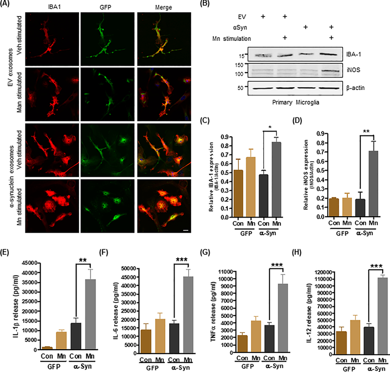
Figure 2: Mn-stimulated exosomes promote neuroinflammatory responses.
(A) Immunofluorescence analysis of primary microglial cells (IBA1; red color) exposed to exosomes (GFP; green color). Hoechst dye stained the nuclei (blue). Magnification, 60X; scale bar, 10 μm. Amoeboid and pseudopodic morphology of primary microglial cells exposed to Mn-stimulated αSyn exosomes was visually assessed (lower images). (B to D) Representative Western blots (B) and densitometry (C and D) assessing IBA-1 and iNOS abundance after exposure to Mn-stimulated αSyn exosomes, as a measure of their potential to promote neuroinflammatory responses in vitro. Data are mean ± SEM (*p≤0.05, **p<0.01 by one-way ANOVA with Tukey’s post-test) of five independent experiments. (E to H) Pro-inflammatory cytokine release upon exosome treatment was quantified using Luminex bead-based cytokine assays. Data are mean ± SEM (**p<0.01, ***p<0.001 by one-way ANOVA with Tukey’s post-test) of four individual experiments performed in 8 replicates.

