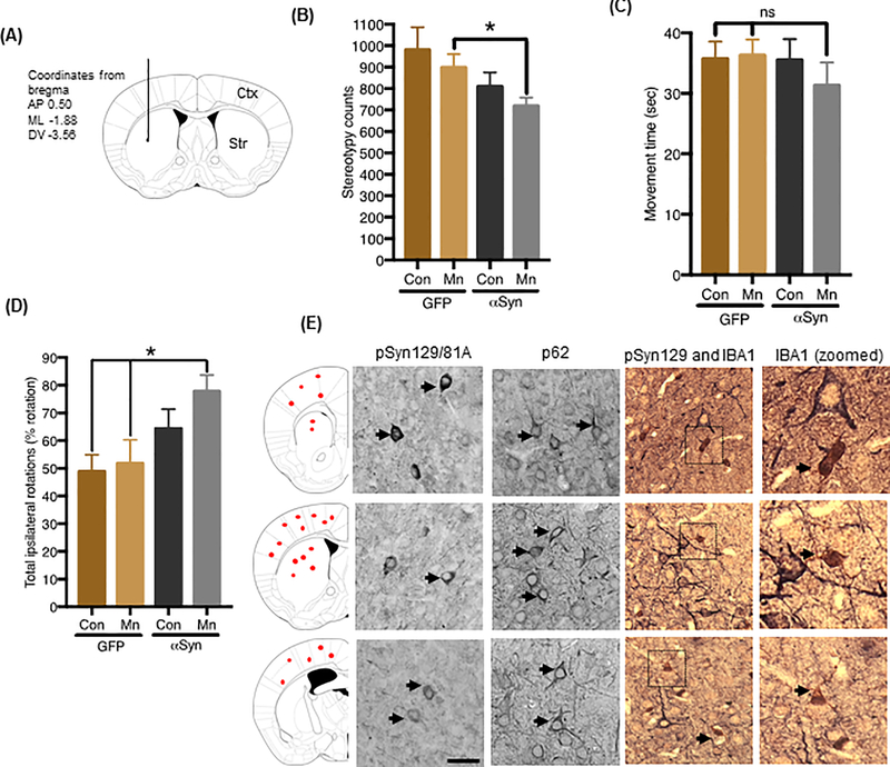
Figure 6: Mn-stimulated αSyn exosomes induce Parkinson-like motor deficits in non-transgenic mice.
(A) Diagram illustrating route and coordinates (mm from bregma) of stereotaxic exosome injections (coronal view). Exosomes were inoculated into the left hemisphere at subdural depths, indicated using a single needle tract. (B and C) Open-field behavior analysis measured using VersaMax apparatus, assessing stereotypy counts (B) and movement time (C) in C57BL/6 mice injected with exosomes isolated from Mn- or vehicle-treated GFP_EV and GFP_Syn cells. Data are mean ± SEM of n ≥12 animals per group; *p≤0.05; ns = not significant by one-way ANOVA with Tukey’s post-test. (D) Immunohistological analysis of phosphorylated αSyn (pSyn129/81A), p62 immunoreactivity, and primary microglial cells (IBA1) in brain tissue from mice injected with Mn-stimulated αSyn exosomes. Corresponding brain regions shown in far-left panel. Magnification, 40X; scale bar, 25 μm. (E) Amphetamine-induced rotation test. The graph shows net scores for ipsilateral rotational asymmetry (number and direction of rotations) induced by amphetamine 180 days post-lesioning in mice receiving vehicle- or Mn-stimulated GFP or αSyn exosomes.

