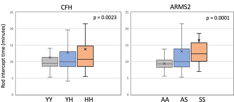
Figure 1.

Box plots showing the distribution of RIT for CFH genotypes and for ARMS2 genotypes with combined data for all eyes in the study (with either normal macular health or with AMD). Associations are adjusted for age and smoking status. The box represents the middle two quartiles of the distribution; the lower whisker, the bottom quartile; the upper whisker, the highest quartile. The median is the horizontal line in each box and the “X” is the mean. Statistical outliers not shown.
