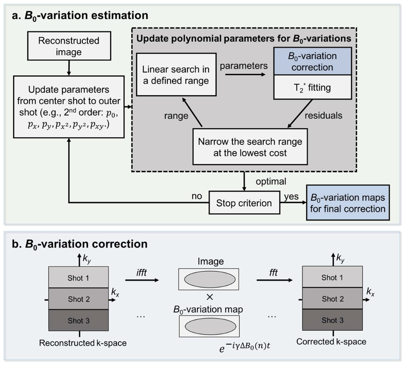
FIG. 3.
An illustration of the proposed self-B0 variation estimation (a) and correction (b). After reconstruction, fully-sampled EPTI-shots can be obtained but with different B0-variations. Polynomial maps are used to estimate these B0-variations assuming they are of sufficient low spatial frequency. To estimate the variation maps, the polynomial parameters are updated iteratively from the center to the outer segments. A linear greedy search algorithm within a selected range is performed for each parameter by selecting the value that gives the minimal residual errors of T2* fitting, assuming that the clean images without artifacts will have minimized residual errors. The estimated B0-variation maps are then used to correct the phase for each segment in the image domain as shown in (b). The corrected segments are combined to obtain the final image series.

