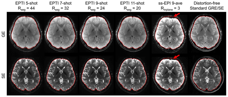
FIG. 6.
Results of in-vivo experiments with different EPTI shots at 1.1×1.1×3 mm3 resolution. Combined GE and SE EPTI images eliminated the severe distortion appeared in ss-EPI (red arrows), and achieved high-SNR artifact-free images under all acceleration factors. The brain boundaries were extracted from distortion-free standard GRE/SE acquisition as shown on the right and overlaid on the EPTI and ss-EPI images for comparison.

