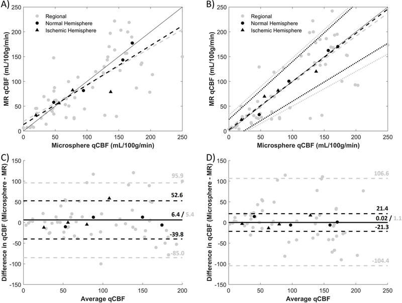
Figure 5.

Correlation and Bland-Altman plots of MR-qCBF vs. microsphere-qCBF with MCAO before (A, C) and after (B, D) delay and dispersion correction. Hemispheric averages are in black and regional averages are in gray. The hemispheres affected with MCAO are shown as triangles, while the contralateral sides are circles. Dashed lines are derived from linear regression analysis, dotted lines represent 95% CI, and a line of unity is shown for reference. An improvement in the hemispheric correlation can be seen after the correction, and the linear regression lines are brought closer to the line of unity. In the Bland-Altman plots (C, D), the bias is represented by a solid line and the dashed lines are the limits of agreement. After correction, the bias for hemispheric and regional are reduced closer to zero.
