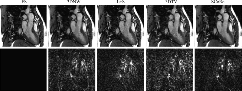
Figure 6:
Reconstructed images from the in vivo study with RU. A representative frame from (Dataset #1) is shown. Results from fully sampled (FS) reference and four different reconstruction methods are shown in the first row. Corresponding error maps after three-fold amplification are shown in the second row. For the images shown, the data were retrospectively undersampled at R = 12.

