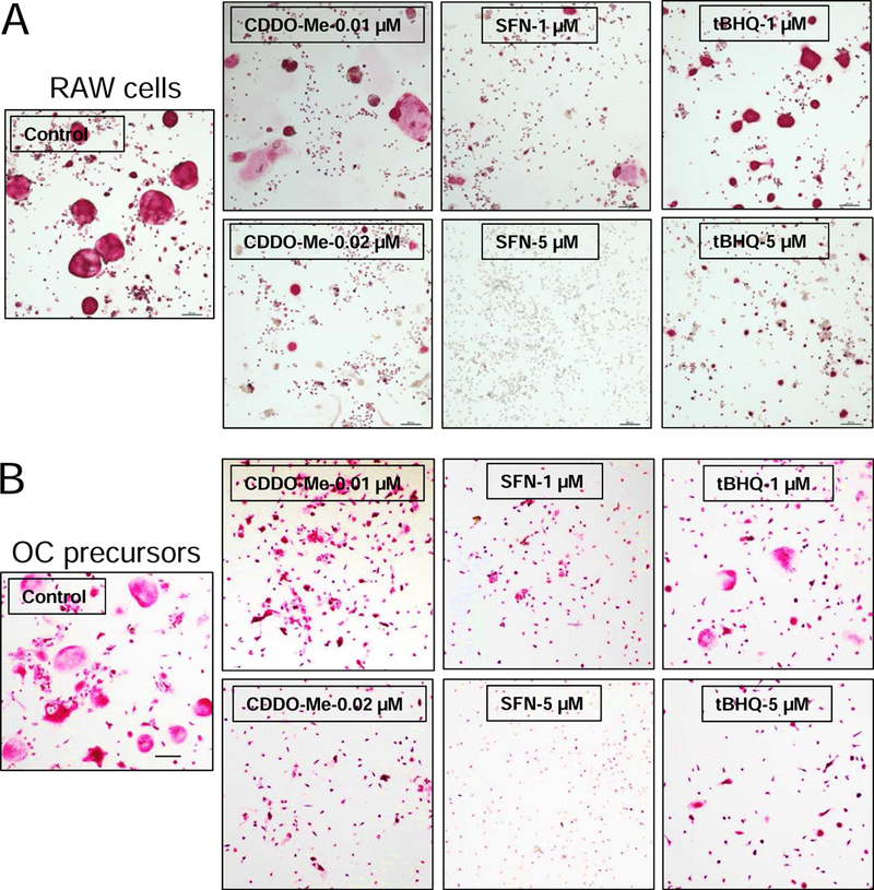
Fig. 1.

CDDO-Me, SFN and tBHQ attenuate the RANKL-induced differentiation in RAW cells and primary osteoclast precursors. (A) RAW cells and (B) primary osteoclast precursors were treated with various concentrations of CDDO-Me, SFN, and tBHQ in the presence of RANKL (10 ng/ml) and M-CSF (30 ng/mL, only applied to primary cells) for 5 days. The cells were then fixed with 10% paraformaldehyde, and stained with TRAP solution. Images were taken using microscope. Representative images are shown in panel. (bar = 200 µm). Control (only treated with RANKL).
