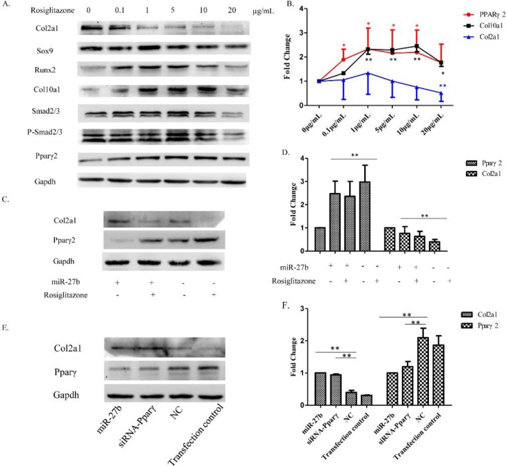
Figure 6. Effect of overexpression or knockdown of Pparγ on the differentiation of chondrocytes.
(A) Resting chondrocytes plated in a monolayer were treated with increasing concentrations of rosiglitazone (0–20 µg/ml). Western blot showing the expression of selected proteins. Gapdh was used as a loading control. (B) Fold-change of Pparγ, Col10a1, and Col2a1 at different concentrations of rosiglitazone compared with untreated resting chondrocytes (n=3, *P<0.05, **P<0.01). (C) Resting chondrocytes were concomitantly treated with miR-27b mimics (75 nM) and rosiglitazone (5 µg/ml). Western blot showing the expression of selected proteins. (D) Semi-quantitation of Western blots showing the fold-change of Col2a1 and Pparγ2 protein expression. Gapdh was used as a loading control (n=3, *P<0.05, **P<0.01). (E) Hypertrophic chondrocytes were transfected with miR-27b mimics (75 nM) and siRNA-Pparγ (50 nM), respectively. Western blot showing the protein level of Col2a1 and Pparγ. (F) Semi-quantitation of Western blots showing the fold-change of Col2a1 and Pparγ2 protein expression. Gapdh was used as a loading control (n=3, *P<0.05, **P<0.01).

