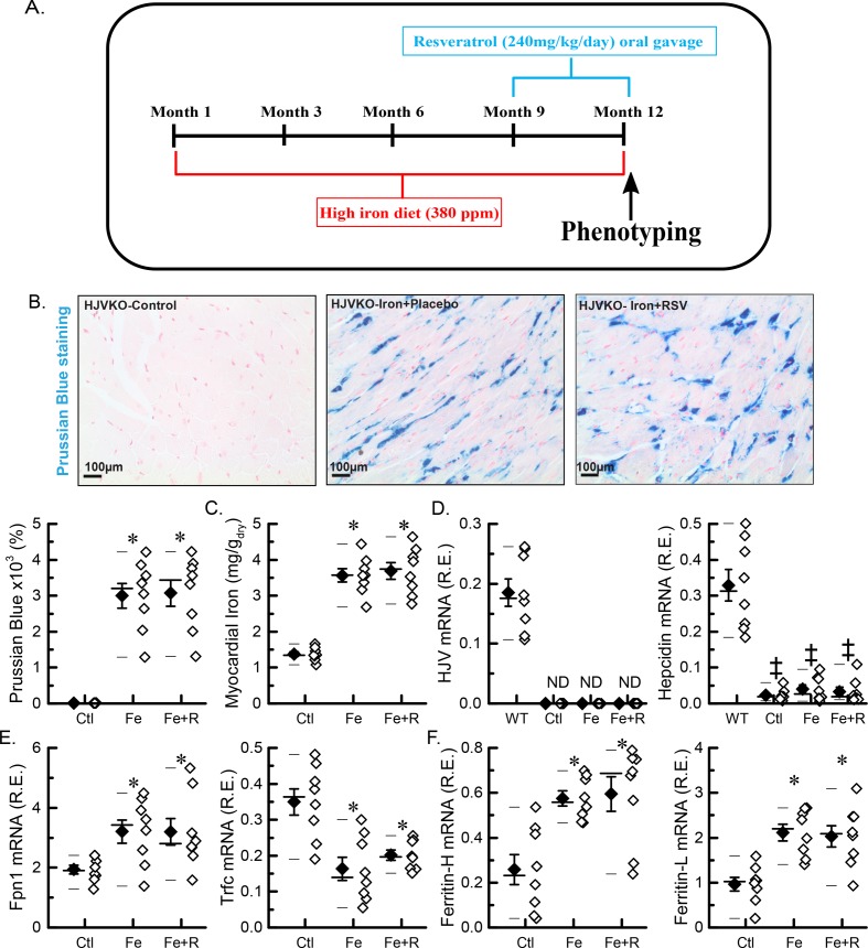
Figure 1. Myocardial iron overload in an aged murine model of iron-overload cardiomyopathy.
(A) Schematic representation of the study design. (B) Representative images for Prussian Blue staining with quantitation (n=8: four hearts; two sections per heart). (C) Total myocardial tissue iron levels (n=8 hearts). (D) Expression of iron metabolic genes: HJV and hepcidin (Hamp). (E) Expression of iron transporting genes: ferroportin (FPN1) and transferrin receptor 1 (Trfc). (F) Expression of iron storage genes: ferritin light (L) chain and ferritin heavy (H) chain. n=8 (hearts) for gene expression analysis (D–F); *P<0.05 compared with the Ctl group. ‡P<0.05 compared with the WT standard group. Abbreviations: Ctl, HJV control; Fe, iron diet; Fe + R, iron diet + resveratrol; ND, not detected.

