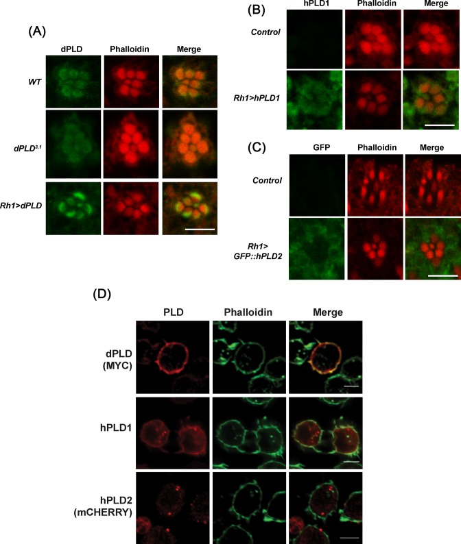
Figure 4. Sub-cellular localization of human PLD proteins in Drosophila cells.
(A) Transverse section (TS) of retinae from WT, dPLD3.1, and Rh1 > dPLD stained with dPLD antibody. Flies were dissected after 0–6 h (day 0) post eclosion. Scale bar: 5 µm. (B) TS of retinae from Control and Rh1 > hPLD1 stained with hPLD1 antibody. Flies were dissected after 0–6 h (day 0) post eclosion. Scale bar: 5 µm. (C) TS of retinae from Control and Rh1 > GFP::hPLD2 stained with GFP antibody. Flies were dissected after 0–6 h (day 0) post eclosion. Scale bar: 5 µm. (D) Confocal imaging showing co-localization of MYC::dPLD (stained with MYC), hPLD1 (stained with hPLD1 antibody raised against N-terminus of the protein) and mCHERRY::hPLD2 (stained for mCHERRY) in S2R+ cells with Alexa Fluor 488-phalloidin (Invitrogen). Scale bar: 5 µm.

