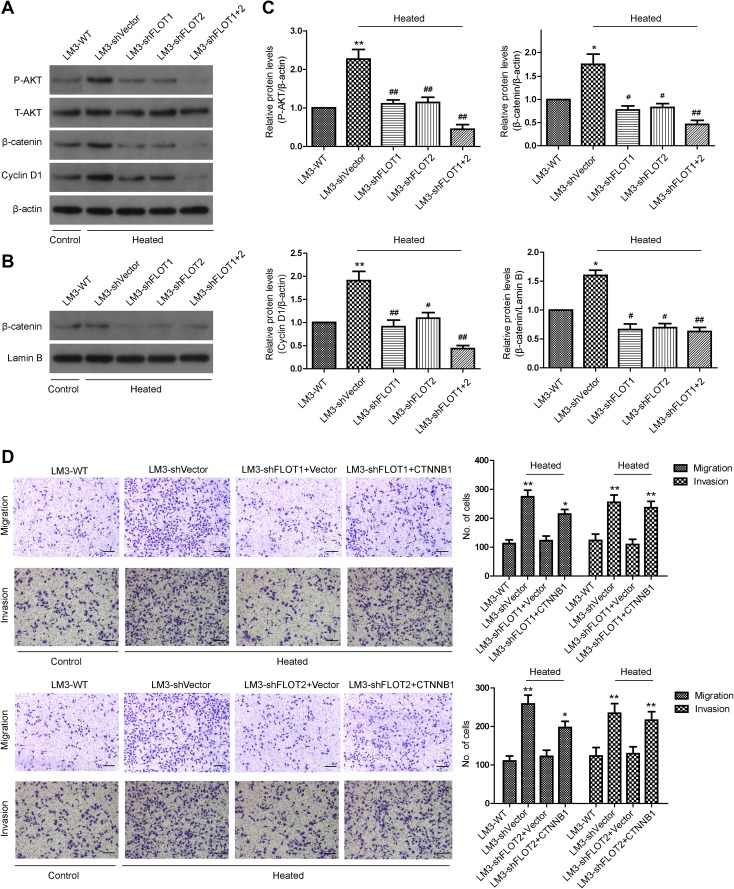
Fig. 5.
The Akt/Wnt/β-catenin pathway plays a vital role in the increased metastatic potential of residual HCCLM3 cells after insufficient RFA. a Western blot analysis of phosphorylated (p)Akt, and total β-catenin and Cyclin-D1 in HCCLM3 cells that were transfected with shVector, shFLOT1, shFLOT2, or shFLOT1 + 2. b Nuclear distribution of β-catenin in HCCLM3 cells was detected by western blot. c Densitometry showing relative changes in FLOT1 and FLOT2 expression. d Representative images of migration and invasion assays of HCCLM3 cells transfected with shVector, shFLOT1, and shFLOT2 ± CTNNB1 were analyzed using transwell assays; scale bar = 100 µm. Data are presented as mean ± SD. Experiments were independently conducted three times; *P < 0.05, **P < 0.01 vs. heat-untreated HCCLM3-WT group; #P < 0.05, ##P < 0.01 vs. heat-treated HCCLM3-shVector group

