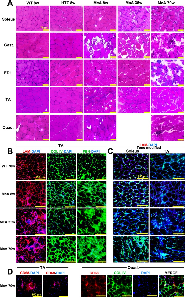
Figure 2.
Histological characterization of McArdle mice. (A) H&E staining of the soleus, gastrocnemius, extensor digitorum longus (EDL), tibialis anterior (TA) and quadriceps muscles from 8-week-old wild-type (WT) and heterozygous (HTZ) and 8, 35 and 70-week-old McArdle (McA) mice. All scale bars correspond to 75 µm. (B) Laminin-DAPI, Col IV-DAPI and FBN-DAPI stains of TA muscles from 8, 35 and 70-week-old McA and 70-week-old WT. All scale bars correspond to 150 µm. (C) Tone modified (Adobe® Photoshop® tone and saturation adjustments were set at −105 and +25, respectively) laminin-DAPI stain of soleus and TA muscles from 70-week-old WT and 8, 35 and 70-week-old McA. Due to the tone modification laminin staining has become dark blue, whereas DAPI staining has become light blue/green and nuclei are much more highlighted. All scale bars correspond to 150 µm. (D) CD68 (pan-macrophage marker)-DAPI staining of TA from 70-week-old McA and CD68, Col IV and DAPI stains of quadriceps from 70-week-old McA mice.

