Figure 3.
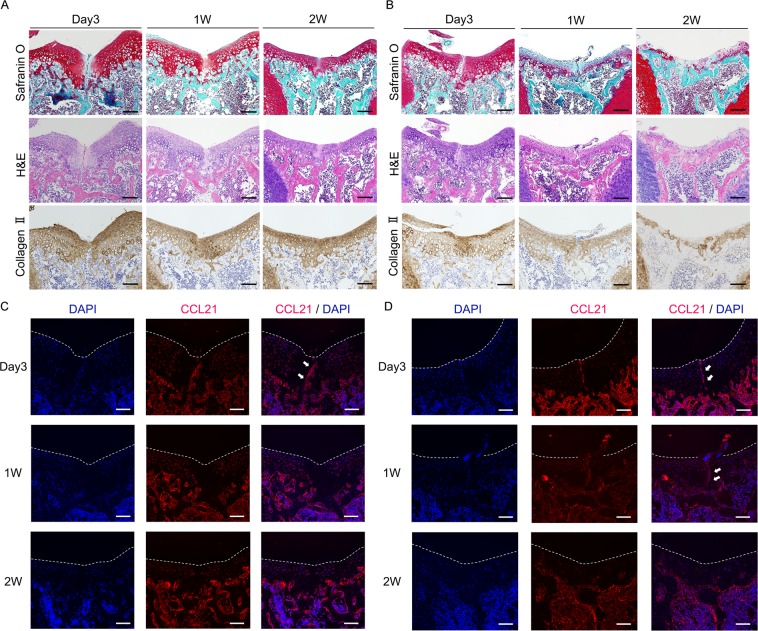
CCL21, CCR7 are expressed at injured sites of juvenile mice in the early phase of post injury. (A) Representative histology in juvenile WT mice with safranin O staining, H&E staining, immunostaining for type II collagen at 3 days, 1 week, and 2 weeks postoperatively. Scale bar: 100 µm. (B) Representative histology in juvenile CCR7−/− mice with safranin O staining, H&E staining, immunostaining for type II collagen at 3 days, 1 week, and 2 weeks postoperatively. Scale bar: 100 µm. (C) Representative images for immunohistochemistry of CCL21 (red) in juvenile WT mice at 3 days, 1 week, and 2 weeks postoperatively. CCL21 was transiently expressed at the injury site in WT mice in the early phase of post injury (white arrow). Scale bar: 100 µm. (D) Representative images for immunohistochemistry of CCL21 (red) in juvenile CCR7−/− mice at 3 days, 1 week, and 2 weeks postoperatively. CCL21 was transiently expressed at the injury site in CCR7−/− mice in the early phase of post injury (white arrow). Scale bar: 100 µm.

