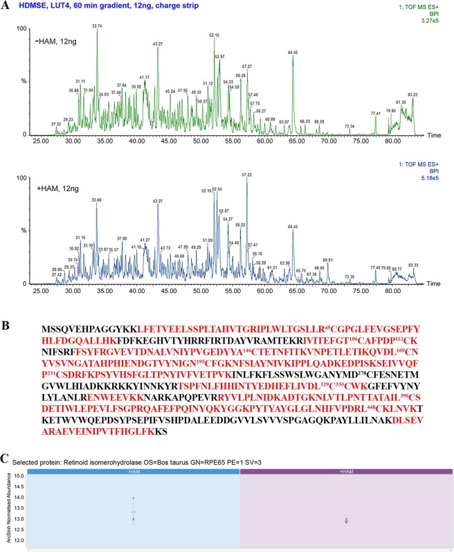
Figure 3.
MS-coupled acyl-labeling of bovine microsome RPE65. (A) Screenshots of LC-HDMSE runs for hydroxylamine-untreated (control) and treated samples from bovine RPE microsomes. (B) MS identified peptides of bovine RPE65 (in red) corresponds to >80% sequence coverage. Numbers in superscript indicate the residue positions of the different cysteines. (C) LC-HDMSE comparison of relative abundance of total RPE65 peptides in the control and HAM-treated bovine microsomes shows that the levels of peptides were similar. The error bars show ± standard error as calculated from the three technical replicate samples.

