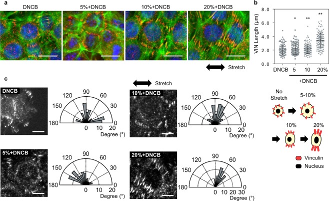
Figure 4.
Redistribution of vinculin (VIN) in HaCaTs in response to chemical and mechanical stimulation. Mechanical stretch applied to cells transduced via focal adhesion in the sites of integrin which initiates downstream signaling. (a) Representative images for vinculin in response to DNCB and mechanical stretch. Scale bars are 20 μm. (b) VIN length as a function of stretch magnitude. P values were calculated using t-test. * and ** mean P < 0.05 and P < 0.005, respectively. (c) Immunostained images of vinculin and its orientation. Focal adhesion was reoriented along 90° (perpendicual to the stretching direction). Scale bars represent 10 µm. n = 105–119 in (b and c).

