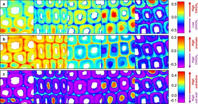
Figure 5.
PCA score images. The PC 1 scores (a) separate the data into pixels with high or low CH/CH2 stretching intensity, while the PC 2 scores (b) separate the data into pixels with high aromatic contribution or high CH/CH2 stretching intensity. The PC 3 scores (c) separate the data into pixels with high contributions from lipophilic extractives or cell wall polymers. Samples from left to right are week 0, 2, 4, 6 and 8. The size of each individual sample image is 70 × 70 μm.

