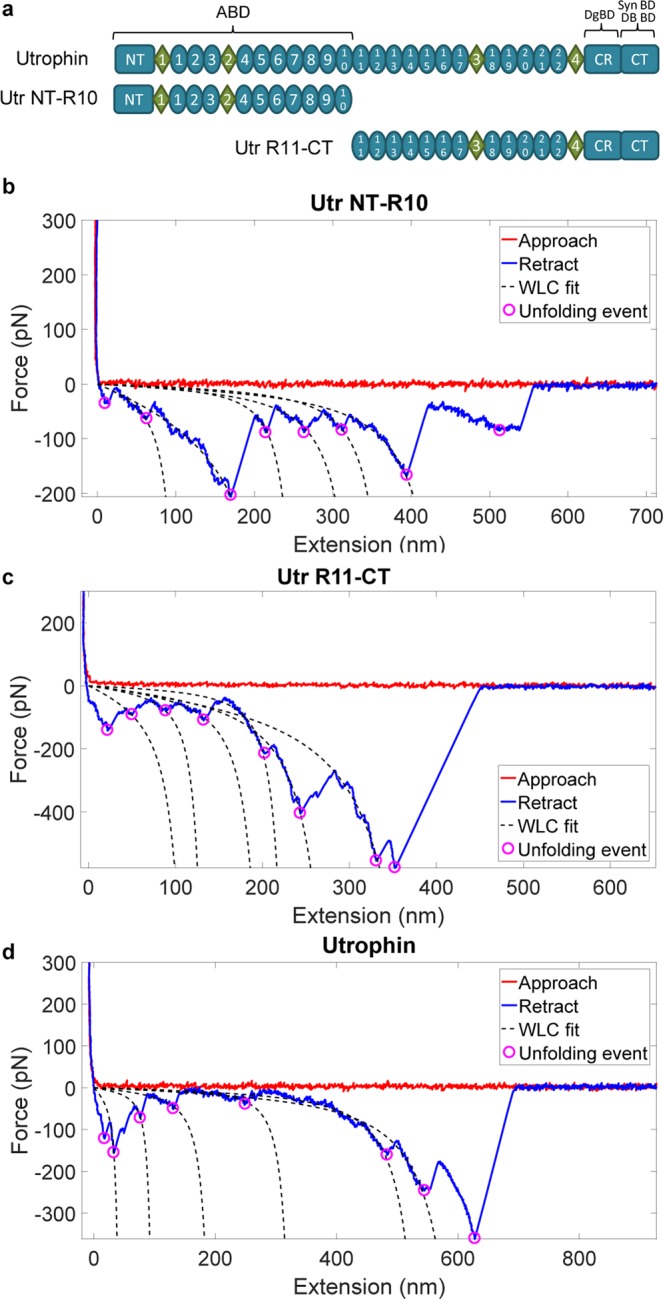
Figure 1.

AFM extension characteristics of utrophin terminal constructs. (a) Schematic of constructs analyzed by AFM. ABD - actin binding domain; DgBD - dystroglycan binding domain; SynBD - syntrophin binding domain; DBBD - dystrobrevin binding domain; NT - N-terminus; CR - cysteine rich domain; CT - C-terminus; ovals - spectrin-like repeats; diamonds - unstructured “hinge” regions. (b–d) Force vs extension representative trace curves for Utr NT-R10 (b), Utr R11-CT (c) and full-length utrophin (d). Approach – force on the cantilever as it approaches the substrate; Retract – force on the cantilever as the molecule is extended; Unfolding events – force minima corresponding to domain unfolding. WLC – worm like chain model fit to the force-extension behavior.
