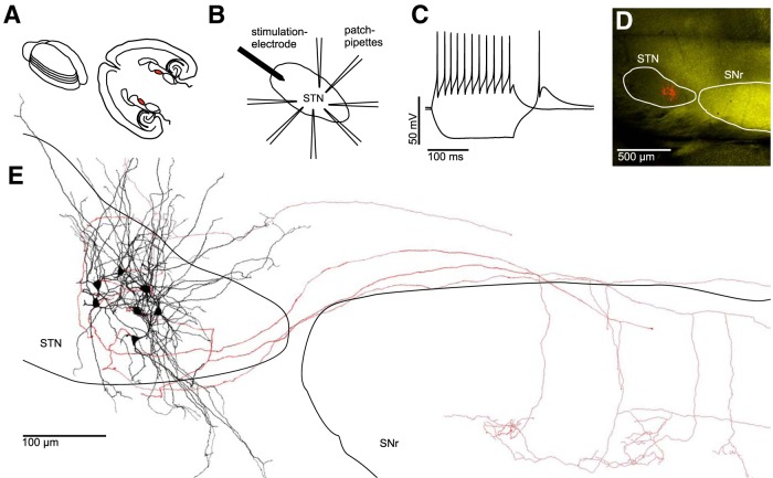
Figure 1.
Electrophysiological and morphological characterization of clusters of neurons within the STN. A, Left, Schematic drawing of an acutely isolated rat brain. Parallel lines indicate orientation of subsequently obtained “horizontal” slices. Right, Anatomical landmarks in a horizontal brain slice with the STN in each hemisphere highlighted in red. B, Experimental paradigm: up to 7 STN neurons were recorded simultaneously. An extracellular stimulation electrode was placed at the rostral tip of the STN to stimulate axons afferent to the cluster of recorded neurons. C, AP pattern of an STN neuron in response to a depolarizing current injection superimposed on a voltage trace in response to a hyperpolarizing current injection, which results in a rebound spike characteristic for STN neurons. D, Fluorescence microscopic image of a horizontal slice obtained from a VGAT-YFP rat (see Materials and Methods) containing both the STN (VGAT-YFP-negative) and the SNr (VGAT-YFP-positive). Red-labeled structures in the STN represent seven simultaneously recorded and biocytin-filled neurons. E, Reconstruction of the same cluster of neurons shown in D. Black represents dendrites and somata. Red represents axons. Note the partially preserved axonal projections to the SNr.

