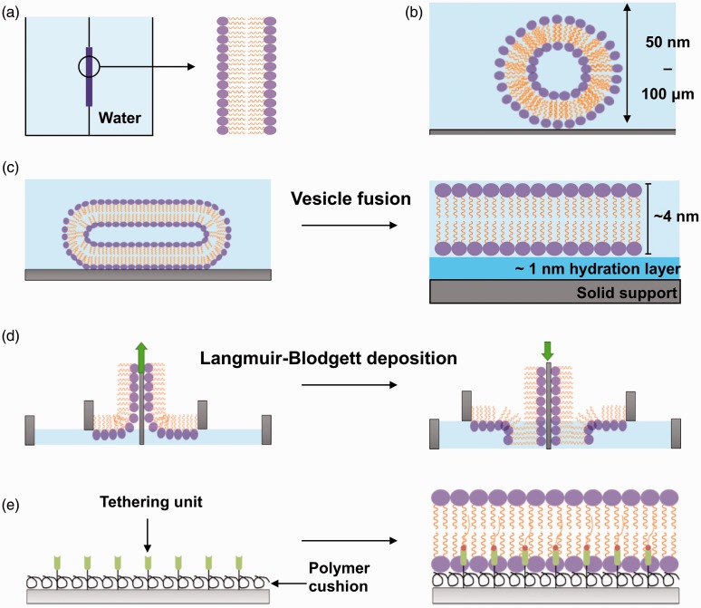
Figure 1.
In vitro approaches used to model the biological membrane. (a) Black lipid membranes are formed when dried down lipids are hydrated with an aqueous medium over an aperture between two aqueous phases. (b) Liposomes (50 nm) or giant uni-lamellar vesicles (100 µm) are formed when dried down lipids are hydrated in an aqueous medium. (c) SLB formation by the vesicle fusion method. Liposomes are incubated onto a hydrophilic surface where they fuse, deform, and spontaneously rupture to form a uniform, continuous, fluid lipid bilayer. (d) SLB formation with the Langmuir-Blodgett technique. Lipid monolayers are formed at the air–water interface when lipids are added to an aqueous medium. This monolayer is transferred onto a hydrophilic solid support twice to form a lipid bilayer. The green arrow indicates the direction the substrate (grey) is moving to pick up another monolayer of lipids. (e) Polymer-supported/tethered lipid bilayers are formed on top of a polymer cushion hydrogel and also tethering units such as nickel NTA for anchoring of proteins. (A color version of this figure is available in the online journal.)

