
Fig. 1.
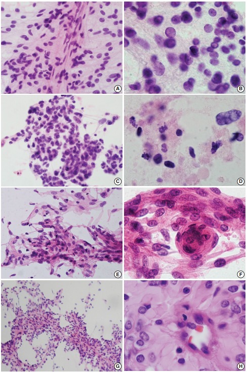
(A, B) Ependymoma. Spindle-shaped tumor cells around thin capillaries. Note fibrillary cytoplasm and occasional nuclear inclusion. (C, D) Glioblastoma. Clusters show ovoid to slightly elongated hyperchromatic nuclei with occasional mitosis. Note necrosis in the extracellular area. (E, F) Fibrous meningioma. Ovoid to spindle cells have round to elongated nuclei with fine chromatin. Note nuclear pseudoinclusion and meningothelial whorls. (G, H) Rhabdoid and papillary meningioma. Papillae have thick collagenous cytoplasm rather than fibrillary features that are commonly found in ependymoma.