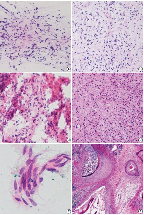
Fig. 2.

Discordant cases. (A, B) Spindle cells with occasional atypical cells are mixed with karyorrhectic debris (A), and glioblastoma is diagnosed in the permanent section (B). (C, D) A tiny focus of round cells shows eosinophilic fibrillary cytoplasm in a bloody background (C), and diffuse astrocytoma is diagnosed in the permanent section (D). (E) Only one cluster of squamoid epithelium is seen in frozen smears. (F) Immature teratoma with neuroepithelium is finally diagnosed in permanent section.
