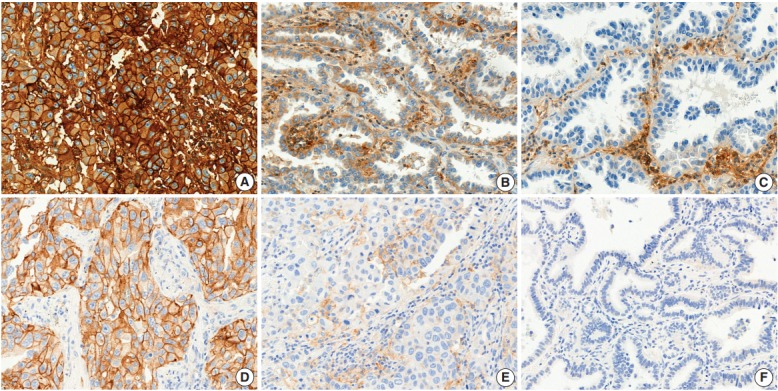
Fig. 1.

Human leukocyte antigen (HLA) class I (A–C) and programmed death-ligand 1 (PD-L1) (D–E) expression in lung adenocarcinoma. (A) Strongly positive (“retained”) staining of HLA class I in tumor cells. (B) Weakly positive staining of HLA class I. (C) Negative staining of HLA class I. (D) ≥50% positive staining of PD-L1 in tumor cells. (E) 1%–49% positive staining of PD-L1. (F) <1% staining of PD-L1.
