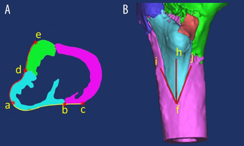
Figure 3.

Three-dimensional computed tomography (3-D CT) reconstruction of the lesser trochanter posterior cortical extension (LTPE) fragment. The posterior cortical extension of the lesser trochanter fragment (LTPE) was measured from the tip (point a) of the LT to the lateral margin of the LT fragment (A, curved line ab). The width of the posterior wall (WPW) was measured from the tip of the LT to the lateral raphé (A, curved line ac). The medial cortical extension of the LT (LTME) fragment was measured from the tip of the LT to the medial margin of the LT fragment (A, curved line ad). The width of the medial wall (WMW) was measured from the tip of the LT to the junction of the anterior and medial cortex (A, curved line ae). The distal cortical extension of the LT (LTDE) fragment was measured from the tip (point f) of the LT fragment to the bottom edge of the LT (B, red line fh). The distal spike angle of the LT (LTDA) fragment was measured as the angle formed by the extension of the two LT fracture lines to the distal femur (B, the angle ifh formed by line if and jf).
