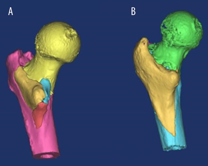
Figure 5.

Three-dimensional computed tomography (3-D CT) reconstruction of the posterior cortical extension of the lesser trochanter (LTPE) fragment. Three-dimensional computed tomography (3-D CT) reconstruction of the 31A2.3 shows several intermediate fragments with an intact greater trochanter (A). 3-D CT images of 31A2.3, which has the LT and greater trochanter and crest in continuity. The tip of the LT fragment is long and sharp (B).
