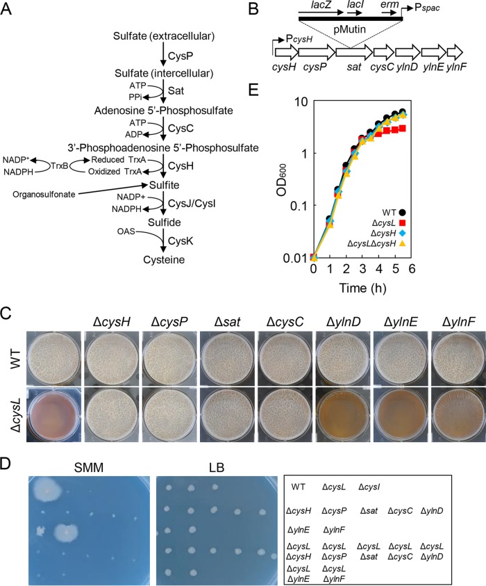
FIG 2.
Null mutations of the cysH operon genes suppress the ΔcysL mutation. (A) Diagram of the cysteine biosynthesis pathway of B. subtilis. The proteins believed to function in the process are shown. (B) Gene organization of the cysH operon. A pMutin insertion into sat is shown above the gene map as an example. (C) Biofilm formation of cysH operon mutants. The wild-type and mutant strains were statically grown for 48 h in 2×SGG medium with 1 mM IPTG. (D) Viability of cysH operon mutants on Spizizen minimal medium (SMM) and LB. Strains were grown at 37°C for 48 h on indicated medium supplemented with 1 mM IPTG. Strain positions on the media are shown in the right panel. (E) Growth profiles of ΔcysH and ΔcysH ΔcysL mutants. Strains were grown at 37°C in 2×SGG medium with vigorous shaking.

