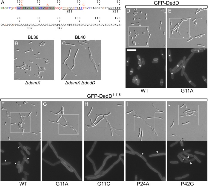
FIG 4.
Identification of residues important for DedD function. (A) DedD residues 1 to 120 are colored according to phylogenetic conservation (percent identity in the Hogenom gene family HOG000279501 [90]); red, 100 to 98%; blue, 98 to 95%; green, 95 to 90%; brown, 90 to 75%; and black, <75%. DedD is a type II bitopic (N-in) inner membrane protein, and its transmembrane domain is shaded in gray. Pertinent substitutions (orange) are indicated immediately above the affected residues. (B and C) DIC images of live BL38 [ΔdamX] (B) and BL40 [ΔdamX ΔdedD] (C) cells after growth for ∼3 mass doublings to an OD600 of 0.5 to 0.6 in M9-maltose. (D to J) Images of live BL40 [ΔdamX ΔdedD] cells carrying a single copy of iBL360 [Plac::gfp-dedD] (D), iBL360(G11A) [Plac::gfp-dedDG11A] (E), iBL345 [Plac::gfp-dedD1–118] (F), iBL345(G11A) [Plac::gfp-dedD1–118, G11A] (G), iBL345(G11C) [Plac::gfp-dedD1–118, G11C] (H), iBL345(P24A) [Plac::gfp-dedD1–118, P24A] (I), or iBL345(P42G) [Plac::gfp-dedD1–118, P42G] (J) integrated in the chromosome. The cells were grown as for panels B and C, but with 250 μM IPTG included in the medium. The panels comprise a DIC image (top) and an FL image (bottom) that corresponds to the boxed area in the DIC image. Bar, 8 μm (DIC images) or 4 μm (FL images). The arrowheads in panels F, I, and J mark examples of the weak accumulation of GFP-DedD1–118 or mutant derivatives at division sites, which are not seen in panels G and H.

