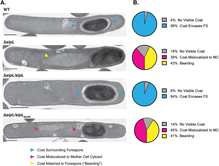
FIG 2.
Loss of SipL’s LysM domain results in coat mislocalization defects. (A) Transmission electron microscopy (TEM) analyses of wild-type 630 Δerm, ΔsipL, and ΔsipL strain complemented with either wild-type sipL or sipL encoding an LysM deletion (sipLΔlysM) after 23 h of sporulation induction. Scale bars, 500 nm. Blue arrowheads mark properly localized coat, i.e., surrounding the entire forespore (FS), whereas pink arrowheads mark coat that has completely detached from the forespore and is found exclusively in the mother cell (MC) cytosol. Yellow arrowheads mark cells where coat appears to be detaching from the forespore but remains partially associated, also known as “bearding” (26). (B) The percentages shown are based on analyses of at least 50 cells for each strain with visible signs of sporulation from a single biological replicate.

