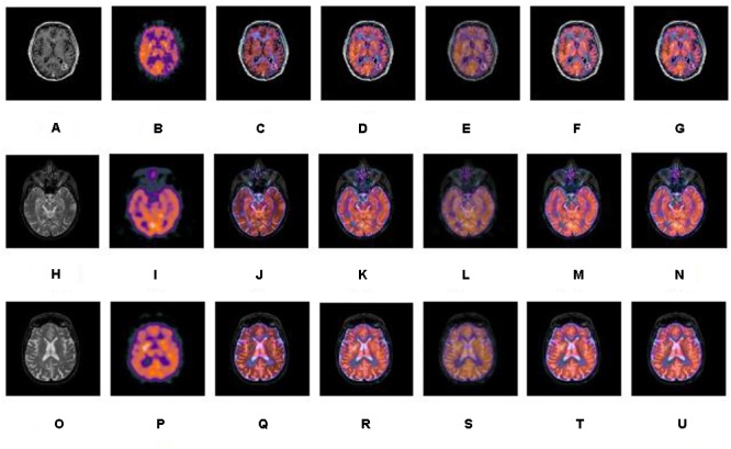
FIGURE 5.

A series of contrasting experiments for normal brain images on fusion images based on different fusion methods (set 1). (A,H,O) are source CT images; (B,I,P) are source SPECT images; (C,J,Q) are fused images based on IHS; (D,K,R) are fused images based on NSCT+FL; (E,L,S) are fused images based on DWT; (F,M,T) are fused images based on the combination of NSCT+PCNN; (G,N,U) are fused images based on the proposed method.
