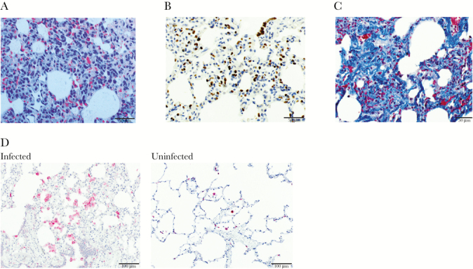
Figure 1.

Lung tissue sections with representative micrographs showing histopathology. (A) Marked type-II pneumocyte hyperplasia alveolar wall thickening and inflammation in a splenectomized monkey, and hemozoin-laden cell infiltration in a hematoxylin and eosin stain-stained section under polarized light. Scale bar = 50 μm. (B) anti-thyroid transcription factor 1 (TTF1) immunohistochemical staining highlights type II pneumocyte hyperplasia in a splenectomized monkey. Type II pneumocytes are indicated by dark brown labeling. Scale bar = 50 μm. (C) Extensive interstitial collagen deposition in alveolar wall stained blue with Masson’s trichrome in the spleen-intact monkey. Scale bar = 50 μm. (D) Numerous immunohistochemically labeled CD163+ cells (fuschin red) infiltrate the alveoli and alveolar walls in the spleen-intact monkey (left) relative to few CD163+ cells in the lungs of an uninfected Saimiri boliviensis monkey (right). Scale bar = 100 μm.
