Figure 2.

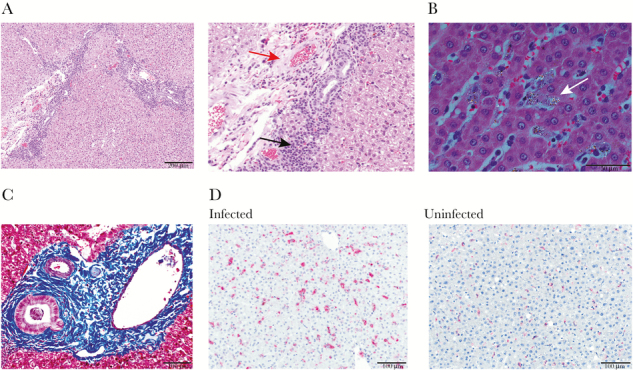
Liver tissue sections of splenectomized monkeys with representative micrographs showing histopathology. (A) Hematoxylin and eosin (H&E)-stained section shows small amounts of collagen deposition and 2 foci of mononuclear periportal infiltrate (left). Scale bar = 200 μm. A zoomed-in image at the same magnification is shown to highlight the collagen deposition (red arrow) and mononuclear infiltrate (black arrow). (B) The H&E-stained section viewed with polarized light shows hemozoin-laden macrophages highlighted by white birefringence with sinusoidal congestion (white arrow). Scale bar = 50 μm. (C) Masson’s trichrome-stained section with collagen deposition in the periportal region shown by deep blue staining. Scale bar = 100 μm. (D) Immunohistochemically stained section showing numerous CD163+-stained cells (fuschin red) and counter stained with hematoxylin in the hepatic parenchyma (left) relative to uninfected Saimiri boliviensis liver tissue (right). Scale bar = 100 μm.
