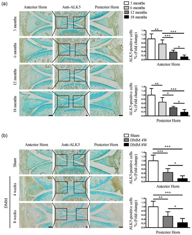
FIGURE 1.
Decreased expression of ALK5 in age-related and OA induced degenerative meniscus anterior and posterior horns. (a) Representative images of IHC staining for ALK5 showed that the number of positive cells in the anterior and posterior horns were strongly reduced with aging. Quantitative data are shown in the right panel (the percentage of positive cells in Cre-negative mice was defined as 1, n = 3 mice per group). (b) Representative images of IHC staining for ALK5 showed that the number of positive cells was gradually reduced in the anterior and posterior horns of mice at 4 and 8 weeks after DMM surgery. Quantitative data are shown in the right panel (the percentage of positive cells in Cre-negative mice was defined as 1, n = 3 mice per group). Scale bar: 100 μm. Data were expressed as the mean ± 95% confidence intervals. *P < 0.05; **P < 0.01; ***P < 0.001. ALK5, activin receptor-like kinases 5; DMM, destabilization of the medial meniscus; IHC, immunohistochemistry; OA, osteoarthritis [Color figure can be viewed at wileyonlinelibrary.com]

