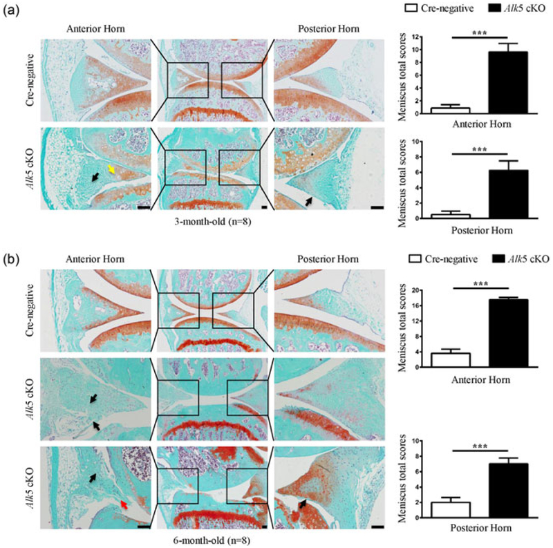
FIGURE 3.
Histological analysis of structural damage in the meniscus of Alk5 cKO mice. (a) Knee joint samples were dissected from 3-month-old mice, and Safranin O/Fast green staining was performed. Representative images showed fibrillation in the superficial zone, inner and outer regions with faint or no Safranin O staining (black arrow) and aberrantly increased hypertrophic chondrocytes (yellow arrow) in menisci of Alk5 cKO mice. Quantitative data are shown in the right panel (n = 8 mice per group). (b) Knee joint samples were dissected from 6-month-old mice, and Safranin O/Fast green staining was performed. Representative images showed more severe fibrillation (black arrow) and disruption of meniscal tissue (red arrow) in meniscus of Alk5 cKO mice. Quantitative data are shown in the right panel (n = 8 mice per group). Scale bar: 100 μm. Data were expressed as the mean ± 95% confidence intervals. *P < 0.05; **P < 0.01; ***P < 0.001. Alk5, activing receptor-like kinases 5 [Color figure can be viewed at wileyonlinelibrary.com]

