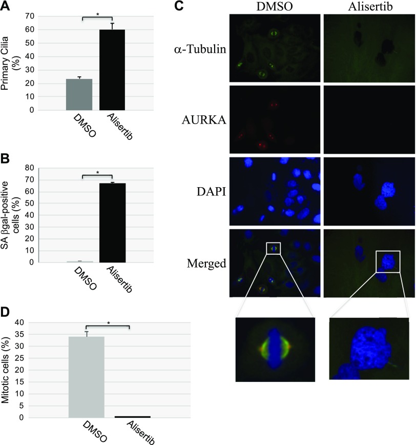
Figure 10.
Alisertib inhibits mitotic spindle formation. HeLa cells were treated for 10 d with alisertib (16 μM). Cells were also treated with DMSO as the control. A, B) Primary cilia formation was quantified by immunofluorescence staining with anti-acetylated α-tubulin (A). Cells were subjected to SA-β-gal activity staining (B). C, D) Cells were synchronized in mitosis by double thymidine block. Immunofluorescence staining was then performed with antibody probes specific for α-tubulin (green), AURKA (red), and DAPI (blue). Representative images (C) and quantification of cells displaying a mitotic spindle (D) are shown. Values represent means ± sem. *P < 0.001 (Student’s t test).

