Figure 14.
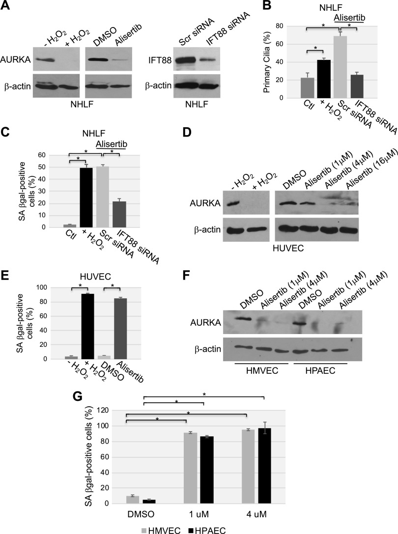
Oxidative stress and alisertib promote down-regulation of AURKA, ciliogenesis, and premature senescence in primary human fibroblasts. A–C) NHLFs were transfected with either scrambled (Scr) or IFT88 siRNA. Cells were then treated with either alisertib (16 μM) for 10 d or with sublethal doses (450 μM) of H2O2 for 2 h, washed, and recovered in complete medium for 10 d. A) cell lysates were subjected to immunoblot analysis with antibody probes specific for either AURKA or IFT88; immunoblot with anti-β-actin IgGs was performed to show equal loading. B) Cells were subjected to immunofluorescence staining with anti-acetylated α-tubulin IgGs to detect primary cilia. Quantification of cells carrying a primary cilium is shown. C) Cells were subjected to SA-β-gal staining. D, E) HUVECs were treated with either sublethal doses of H2O2 (450 μM) for 2 h and recovered in complete medium for 7 d or alisertib (1, 4 and 16 μM) for 10 d. D) AURKA protein expression was determined by immunoblot analysis with an AURKA-specific antibody probe; immunoblot with anti-β-actin IgGs served as the control. E) Cells were subjected to SA-β-gal activity staining. F, G) HPAECs and human lung microvascular endothelial cells were treated with alisertib (1 and 4 μM) for 10 d. F) Cell lysates were subjected to immunoblot analysis, with antibody probes specific for AURKA; immunoblot with anti-β-actin IgGs was performed as the control. G) Cells were subjected to SA-β-gal activity staining. Values represent means ± sem. *P < 0.001 (Student’s t test).

