Figure 5.
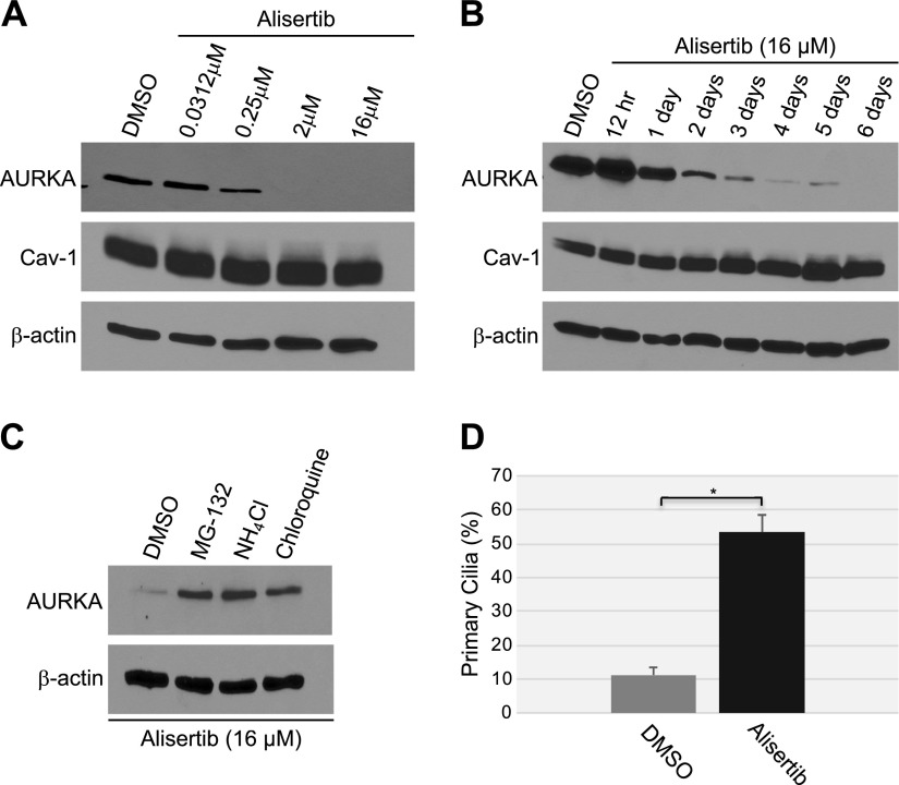
Alisertib induces degradation of AURKA and ciliogenesis. A) WI-38 human diploid fibroblasts were treated with different concentrations of alisertib (0.0312, 0.25, 2 and 16 μM) for 6 d. B) WI-38 fibroblasts were treated with 16 μM alisertib for different times (12 h and 1, 2, 3, 4, 5 and 6 d). Treatment with DMSO was used as the control. A, B) Cells were collected, and the expression levels of AURKA and caveolin (Cav)-1 were determined by immunoblot analysis with anti-AURKA and anti-caveolin-1 IgGs. Immunoblot with anti-β-actin IgGs was performed to show equal loading. C) WI-38 fibroblasts were treated with 16 μM alisertib for 6 d in the presence of MG-132 (0.6 μΜ), ammonium chloride (NH4Cl; 10 mM), or chloroquine (50 μΜ). Treatment with DMSO served as the control. Cells were collected, and cell lysates were subjected to immunoblot analysis with antibody probes specific for AURKA and β-actin (to show equal loading). D) WI-38 cells were treated with 16 μM alisertib for 6 d. Cells were then subjected to immunofluorescence analysis using anti-acetylated α-tubulin IgGs. Quantification of the staining is shown. Values represent means ± sem. *P < 0.001 (Student’s t test).

