Figure 6.
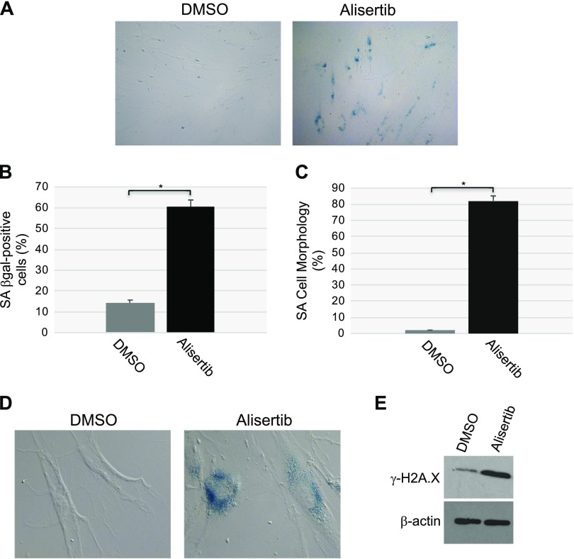
Treatment with alisertib promotes accumulation of cells displaying SA-β-gal activity, senescent cell morphology, and elevation of phosphorylated H2A.X. A, B) WI-38 human diploid fibroblasts were treated with 16 μM alisertib for 10 d. DMSO-treated cells were used as the control. Cells were then subjected to SA-β-gal activity staining. Representative low-magnification images (A) and quantification of the staining (B) are shown;. C, D) WI-38 human diploid fibroblasts were treated with 16 μM alisertib for 10 d. Treatment with DMSO was used as control. Cells showing senescence-associated cell morphology were identified. Quantification of the percentage of cells showing senescence-associated cell morphology (C) and representative high-magnification images (D) are shown. D) SA-β-gal activity staining is also shown. E) WI-38 human diploid fibroblasts were treated with 16 μM alisertib for 10 d. DMSO-treated cells were used as the control. Cells were collected, and cell lysates were subjected to immunoblot analysis with an antibody probe specific for phosphorylated histone H2A.X (γ-H2A.X). Immunoblot analysis with anti-β-actin IgGs was performed to show equal loading. Values represent means ± sem. *P < 0.001 (Student’s t test).

