Figure 9.
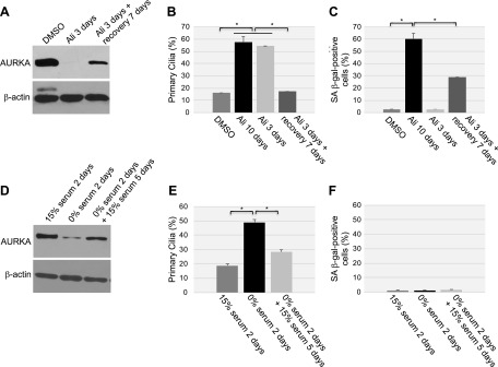
Transient down-regulation of AURKA temporarily prevents the disassembly of the primary cilium but does not promote cellular senescence. A–C) WI-38 fibroblasts were continuously treated with DMSO or alisertib (16 μM) for 3 or 10 d. Cells were also treated with alisertib (16 μM) for 3 d, washed, and cultured for an additional 7 d in the absence of alisertib. A) Cell lysates were subjected to immunoblot analysis with an antibody probe specific for AURKA. Immunoblot analysis using anti-β-actin IgGs was performed to show equal loading. B) Primary cilia formation was quantified by immunofluorescence staining with an antibody probe specific for acetylated α-tubulin. C) Cellular senescence was quantified after cells were subjected to SA-β-gal activity staining. D–F) WI-38 cells were cultured in either serum-containing or serum-free medium for 2 d. Serum-starved cells were then cultured for an additional 5 d in the presence of serum. D) AURKA protein expression was determined by immunoblot analysis using anti-AURKA IgGs. Immunoblot analysis with anti-β-actin IgGs was performed as control. E) Primary cilia formation was quantified by immunofluorescence staining with an antibody probe specific for acetylated α-tubulin. F) Cells were subjected to senescence-associated (SA)-β-galactosidase activity staining. Values represent means ± sem. *P < 0.001 (Student’s t test).

