Figure 4.
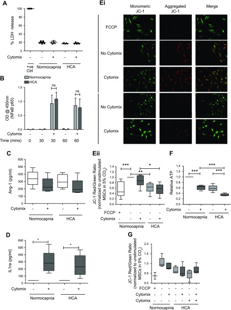
HCA induces mitochondrial dysfunction in MSCs. A) Viability of MSCs was unaffected by HCA at 24 h when assessed by measurement of LDH release (n = 2 per group). B) The degree of cytomix-induced NF-κB activation in MSCs was unaffected by HCA at 30 or 60 min when assessed based on the relative amount of active p65 subunits present in nuclear extracts obtained from MSCs (n = 5 per group for 0 and 60 min; n = 4 per group for 30 min). C) MSC secretion of the soluble mediator Ang-1 was not altered by HCA at 24 h when quantified by ELISA (n = 5 per group). D) The quantity of the soluble mediator IL-1ra secreted by MSCs was also unaffected by HCA at 24 h (n = 5 per group). Ei, ii) Staining with the mitochondrial membrane potential indicator JC-1 revealed significant attenuation of HPMEC mitochondrial membrane potential by HCA in both the presence and absence of cytomix stimulation at 24 h. Ei) Representative images. Eii) The JC-1 red (aggregate)/green (monomer) ratio was quantified in ImageJ and presented relative to unstimulated cells in normocapnia (n = 5 per group). F) HCA attenuated ATP production by MSCs at 24 h (n = 5 per group). G) Buffering medium pH to that of normocapnia using 0.02 M NaHCO3 did not alter the effect of HCA on mitochondrial membrane potential in MSCs at 24 h (n = 3 per group except FCCP and all groups not stimulated with cytomix in HCA, which are n = 2). Images were taken on an EVOS FL Auto epifluorescence microscope. Ctrl. Control; ns, not significant; OD, optical density; +ve, positive control. Scatter plots show mean and sd, and box and whisker plots show median, first and third quartiles, and maximum and minimum values. Original magnification, ×20. *P < 0.05, **P < 0.01, ***P < 0.001.

