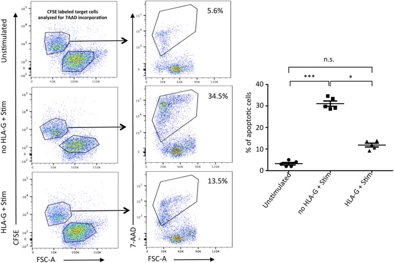
Figure 9.
HLA-G dimer inhibits cytotoxicity of CD8+ T cells. PBMCs from HVs (n = 5/group) were stimulated with ConA-IL-2 as described in Materials and Methods. Data from flow cytometry–based killing assay are shown. In the representative flow cytometry dot plots (left panels) K562 target cells are shown as CFSE-positive cells and the effector PBMCs are CFSE-negative populations. CFSE-positive target cells were gated and analyzed for 7-AAD incorporation as an early indicator of cell death (right panels). Graphical representation indicating percent of apoptotic cells per group. Representative from 1 of 3 separate experiments is shown. N.s., not significant. Data presented as means ± sd. *P < 0.05, ***P < 0.001.

