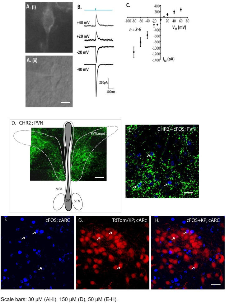
Fig 4. AAV-ChR2 delivery to PVN neurons and optogenetic responses in the ARC.
AAV-ChR2 was delivered by stereotaxic injection into the PVN and 8 weeks later, brain slices were stimulated by blue light to evaluate direct synaptic connectivity between PVN neurons and Kiss1 neurons in the arcuate. A. Expression of AAV-ChR2-GFP in PVN neurons 8 weeks after stereotaxic injection into PVN (i) GFP (ii) Light. B. Example responses of a single PVN neuron to brief light stimulation (blue; 480 nm, 10 ms duration) at different membrane potentials. C. Current voltage relationship for light activated responses in PVN neurons. D. A composite of confocal images indicating neurons expressing ChR2 within the medial posterodorsal portion of the PVN. E. Robust expression of the AAV-ChR2 vector was confirmed by GFP immunofluorescence and c-FOS induction (blue, arrowed) in the PVN after light stimulation (Fig 4E). F. c-FOS expression in the caudal ARC after light stimulation. G. Kiss1 neurons in the caudal ARC visualized by TdTomato expression. H. Merged image showing limited activation of c-FOS expression in Kiss1 neurons. ARC, arcuate nucleus; PVN, paraventricular nucleus; ac, anterior commissure. MPA, medial preoptic are; PVNmpd, paraventricular nucleus medial posterodorsal; SCN, suprachiasmatic nucleus; 3V, 3rd ventricle. Scale bars: 30 μM (Ai-ii), 100 μM (D), 50 μM (E-H).

