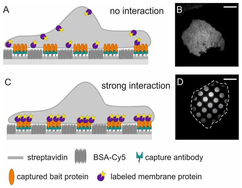
Figure 1. Principle of Protein Micropatterning in the plasma membrane.
(A) Sketch and (B) TIRF image of a cell grown on a micropatterned substrate. Bait antibody is arranged in a regular pattern of 3 μm sized dots with 3 μm interspaces. The bait protein (unlabeled) reorganizes according to the antibody patterns, but the fluorescently labeled prey protein is distributed homogeneously in the plasma membrane, indicating no interaction between bait and prey protein. Scale bar is 7 μm. (C,D) As in (A,B), but here the prey protein interacts strongly with the bait protein and localizes according to the bait patterns. The cell outline is indicated by a dashed white contour line.

