Figure 5.
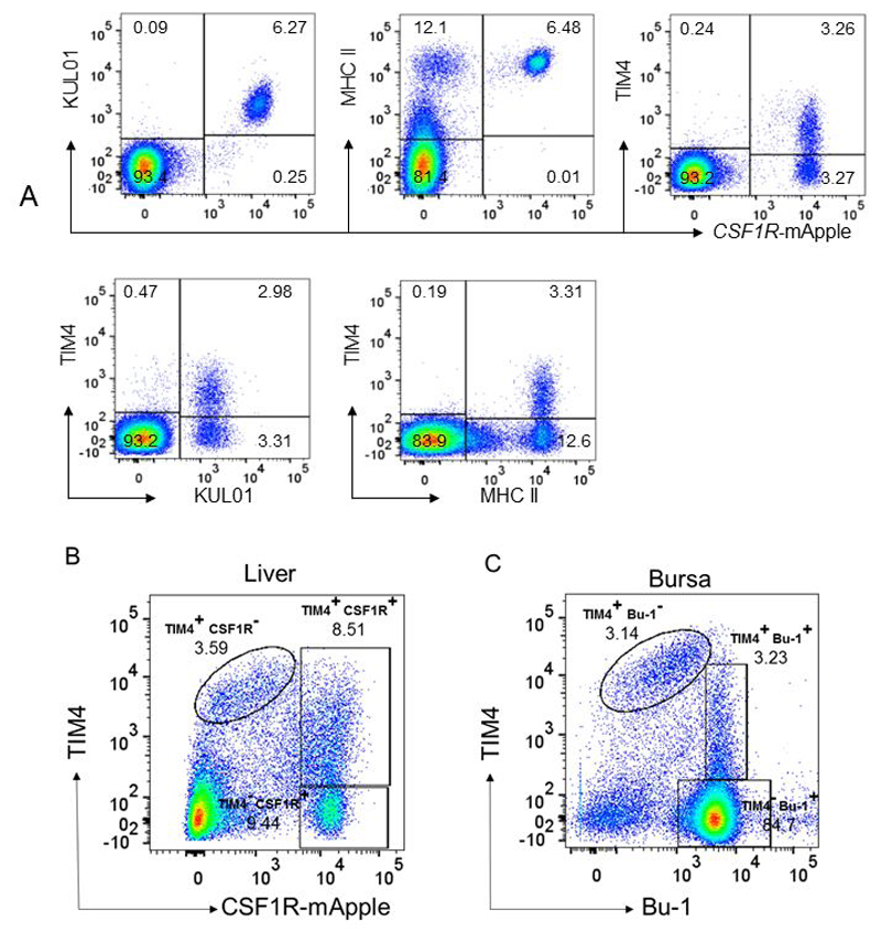
FACS separation of CSF1R-mApple+ and TIM4+ cells in blood, liver and bursa for RNAseq analysis.
A) Pooled blood leukocytes from CSF1R-mApple birds (n = 6) were labelled with anti-KUL01, MHCII or TIM4 as indicated. Note the uniform expression of KUL01 and MHCII on CSF1R-mApple+ cells, and heterogeneous expression of TIM4. B) Pooled non-parenchymal cells isolated from liver of CSF1R-mApple birds as described in Materials & Methods (n = 7). Note the separation of TIM4+CSF1R-mApple- cells (Kupffer cells) from two populations CSF1R-mApple+ cells differing in expression of TIM4. C) Pooled isolated cell populations from bursa of Fabricius (n = 7) were double stained for TIM4 and Bu-1 as described in Materials and Methods. Note the presence of a population of TIM4+, Bu-1- cells (macrophages) and two populations of Bu-1+ cells distinguished by TIM4 expression.

