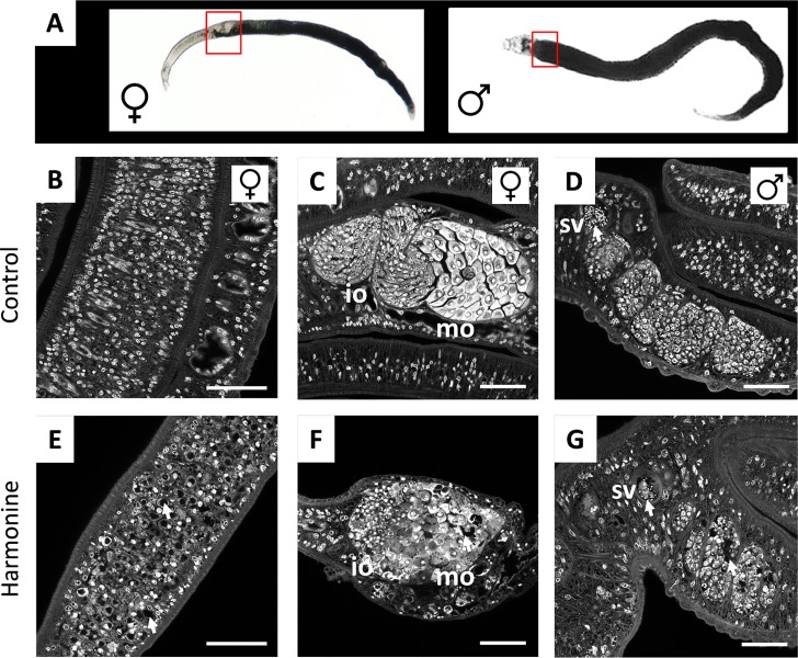
Fig 6. Effect of harmonine on gonadal tissue structure.
For CLSM-analysis of gonadal tissues, S. mansoni couples were cultured for 72 h with 10 μM harmonine (E-G) or solvent as a control (B-D), and stained with carmine red. (A) Bright-field microscopic images indicating the localization of ovary and testes in worms. (B, E) Confocal images showing part of the intact vitellarium of a control female (still paired with a male) (B) compared to the porous appearance of the vitellarium after harmonine treatment (E). (C, F) Well-defined immature (iO) and mature (mO) parts of a control ovary (C), compared to the disintegrated structure of an ovary in a harmonine-treated female (F). (D, G) Seminal vesicle (sv) filled with spermatozoa, and testes lobes filled with spermatogonia of a control male (D), compared with the gonad of a harmonine-treated male with reduced number of spermatozoa and partially disintegrated lobes (arrows) (G). Scale bar: 50 μm.

