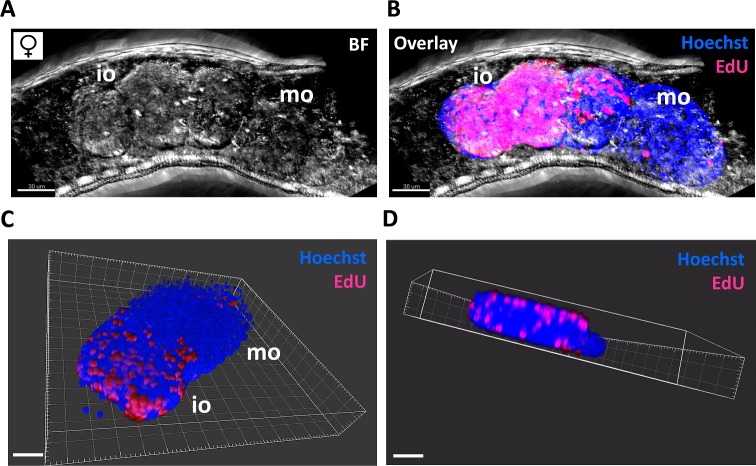
Fig 7. 3D reconstruction of S. mansoni ovary and stem cell localization.
Couples were cultured for 24 h with EdU. Females were separated from males, stained with Hoechst, and z-stacks of the ovary and surrounding tissue were acquired by CLSM. Z-stacks were processed with the IMARIS software to release the ovary from the surrounding tissue and to quantify the number of EdU- and Hoechst-positive cells. (A) Representative z-stack showing the immature part (iO) and mature part (mO) of an ovary within a female worm in bright field (BF). (B) The same z-stack with an overlay of the BF image and the in silico-released ovary. Proliferating, EdU-positive stem cells are depicted in pink, Hoechst-positive cells in blue. (C, D) Still images of a video animation of a 3D reconstructed ovary (see supplementary video, S1 Movie). (D) shows the preferential localization of stem cells at the edges of the immature part of the ovary. Scale bar: 30 μm (A, B) or 40 μm (C, D).

