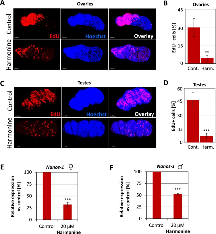
Fig 8. Reduction of gonadal stem cell proliferation upon harmonine treatment.
S. mansoni couples were treated with 20 μM harmonine in vitro for 6 days. As control, an equivalent volume of the solvent DMSO was used. EdU was added for the last 24 h of culture, worm couples were separated and images were processed using the IMARIS software as described in the text and Fig 6. (A, B) Representative images (A) and summary of the analyses of four ovaries (B) from either control or harmonine-treated females. A significant reduction of stem cells was found after treatment, expressed as percentage of EdU-positive per Hoechst-positive cells. (C, D) Representative images (C) and summary of four testes (C) from control and harmonine-treated males showing a significant reduction of the percentage of stem cells following treatment. Scale bar: 30 μm. (E, F) Expression of the gonadal stem cell marker nanos-1 (Smp_055740) in females (E) and males (F) after treatment with 20 μM harmonine compared to control worms as determined by qPCR. The expression in control worms was set to 100%. Summary of two experiments. An unpaired t-test was performed to reveal significant differences (** p<0.01, *** p<0.001).

